


Attraverso la rielaborazione di un software nato per il monitoraggio della crosta terrestre, i ricercatori sono giunti a svilupparne un tipo in grado di rielaborare le immagini delle radiografie in immagini TAC.
Lo sviluppo della ricerca e delle alte tecnologie realizzato dall’INGV trova applicazione anche in molte altre branche della ricerca scientifica. È il caso dello studio “Pipeline for Advanced Contrast Enhancement (PACE) of Chest X-ray in Evaluating COVID-19 Patients by Combining Bidimensional Empirical Mode Decomposition and Contrast Limited Adaptive Histogram Equalization (CLAHE)” recentemente pubblicato sulla rivista ‘Sustainability’ di MDPI, condotto in collaborazione con l’Università di Messina e l’Università di Catania, per lo sviluppo di un applicativo software chiamato “PACE” prodotto per offrire un importantissimo supporto ai radiologi nella diagnosi e nella cura delle patologie polmonari gravi come quelle causate dal COVID-19.
Infatti, è venuta in aiuto alla diagnostica per immagini applicata ai pazienti affetti da patologie polmonari quella normalmente utilizzata dall’INGV per caratterizzare lo “stato di salute” della crosta terrestre.
“L’analogia tra l’interno della Terra e interno dei polmoni può apparire alquanto audace”, afferma Massimo Chiappini, ricercatore dell’INGV e coautore dell'iniziativa. “Tuttavia questa ricerca nasce proprio dall’intuizione di utilizzare su immagini mediche le stesse tecniche di trattamento delle immagini che utilizziamo normalmente per la rappresentazione del sottosuolo nelle aree soggette a rischio sismico, vulcanico o ambientale”.
È noto infatti che per i pazienti affetti da gravi patologie polmonari come, negli ultimi tempi il COVID-19, la valutazione radiologica di lesioni polmonari è indispensabile sia per il monitoraggio dell’evoluzione della malattia sia per valutare la risposta alle specifiche terapie. Tuttavia, quest’attività è resa complessa dal fatto che i pazienti, specialmente nelle fasi acute della malattia, non sono collaborativi e/o si trovano in terapia intensiva. In tali situazioni, inoltre, i radiogrammi sono effettuati spesso con strumenti radiografici portatili che, spesso, producono immagini artefatte che ne riducono la leggibilità.
Pertanto, il software PACE, sviluppato dal team multidisciplinare dei ricercatori dell’INGV, dell’Università di Messina (guidato dal prof. Giovanni Finocchio del Dipartimento di Scienze Matematiche e Informatiche, Scienze Fisiche e Scienze della Terra (MIFT) e dal prof. Giuseppe Cicero del Dipartimento di Scienze biomediche, odontoiatriche e delle immagini morfologiche e funzionali) e dell’Università di Catania (guidato dai proff.i Giulio Siracusano e Aurelio La Corte), è stato ideato per risolvere questi problemi di rappresentazione grafica ottimizzando al massimo il contrasto delle immagini radiografiche del torace.
Ad oggi, vista l’urgenza, i medici l’hanno applicato alle immagini raccolte sui pazienti COVID-19 del Policlinico Universitario “G. Martino” di Messina: con PACE è migliorata significativamente la lettura del radiogramma da parte del radiologo. L’algoritmo, infatti, combina lo stato dell’arte di applicativi numerici di elaborazione delle immagini, quali la decomposizione empirica bi-dimensionale, il filtro omomorfico e l’equalizzazione adattiva dell'istogramma in modo opportuno.
“Dal punto di vista clinico” - afferma il prof. Gaeta del Dipartimento di Scienze biomediche, odontoiatriche e delle immagini morfologiche e funzionali dell’Ateneo di Messina, “è stato fondamentale verificare che le informazioni aggiuntive prodotte da PACE fossero reali. Per far questo, sono state effettuate e confrontate congiuntamente le radiografie del torace e quelle delle TAC tradizionali: il grande successo è stato quello di verificare che le lesioni aggiuntive che il software PACE rilevava nelle semplici immagini radiografiche fossero tutte confermate dalle TAC”.
“La ricaduta del software ideato da INGV e dalle Università siciliane è rilevantissima in ambito sociale” afferma Massimo Chiappini. “Tra i vantaggi, infatti, oltre la evidente riduzione dei costi e di tempi derivante dalla non indispensabilità dei macchinari per la TAC per avere identici risultati diagnostici utili, con l’uso di PACE è sufficiente effettuare un solo intervento sul paziente per l’esame radiografico con un minor rischio di diffusione di malattie virali anche tra gli operatori sanitari come nel caso del COVID-19. Inoltre, questa tecnologia offre la possibilità di applicarla anche in condizioni limite dove l’accesso alla diagnostica TAC non è agevole sia per l’alto numero di degenti interessati sia per i costi della macchina stessa che, nelle aree economicamente poco sviluppate, quali l’Africa ed il Sud America, rappresenta una strumentazione proibitiva”.
Dato l’alto interesse riscontrato in ambito medico, tutti i risultati della ricerca sono a stati messi a disposizione della comunità scientifica liberamente.
Link:
---
From the monitoring of seismic and volcanic territories to medical therapies PACE: the software that replaces the CT scan, is born
Through the reworking of a software created for monitoring the earth's crust, the researchers have come to develop a type capable of reworking the images of the radiographs into CT images.
The development of research and high technologies carried out by INGV also finds application in many other branches of scientific research. This is the case of the study "Pipeline for Advanced Contrast Enhancement (PACE) of Chest X-ray in Evaluating COVID-19 Patients by Combining Bidimensional Empirical Mode Decomposition and Contrast Limited Adaptive Histogram Equalization (CLAHE)" recently published in the MDPI 'Sustainability' journal. The study was conducted in collaboration with the University of Messina and the University of Catania, for the development of a software application called "PACE" produced to offer extremely important support to radiologists in the diagnosis and treatment of severe lung diseases such as those caused by COVID-19.
In fact, the diagnostic imaging normally used by INGV to characterize the “state of health” of the earth's crust has come to the aid of diagnostic imaging applied to patients suffering from pulmonary diseases.
“The analogy between the interior of the Earth and the interior of the lungs can appear quite bold", says Massimo Chiappini, researcher at INGV and co-author of the initiative. "However, this research arises precisely from the intuition of using the same image processing techniques on medical images that we normally use for the representation of the subsoil in areas subject to seismic, volcanic or environmental risk”.
In fact, it is known that for patients suffering from serious lung diseases such as, in recent times, COVID-19, the radiological evaluation of lung lesions is essential both for monitoring the evolution of the disease and for assessing the response to specific therapies. However, this activity is made complex by the fact that patients, especially in the acute phases of the disease, are not cooperative and / or are in intensive care. Furthermore, in such situations, radiograms are often performed with portable radiographic instruments which often produce artificial images that reduce their readability.
Therefore, the PACE software, developed by the multidisciplinary team of researchers from INGV, of the University of Messina (led by Prof. Giovanni Finocchio of the Department of Mathematical and Computer Sciences, Physical Sciences and Earth Sciences (MIFT) and by Prof. Giuseppe Cicero of the Department of Biomedical, Dental and Morphological and Functional Imaging Sciences) and of the University of Catania (led by Profs Giulio Siracusano and Aurelio La Corte), was designed to solve these graphical representation problems by maximizing the contrast of chest radiographic images.
To date, given the urgency, doctors have applied it to the images collected on COVID-19 patients at the University Hospital “G. Martino” in Messina: with PACE, the radiologist's reading of the radiogram significantly improved. The algorithm, in fact, combines the state of the art of numerical image processing applications, such as the two-dimensional empirical decomposition, the homomorphic filter and the adaptive equalization of the histogram in an appropriate way.
“From a clinical point of view” - says prof. Gaeta of the Department of Biomedical, Dental and Morphological and Functional Imaging Sciences of the University of Messina, “it was essential to verify that the additional information produced by PACE was real. To do this, chest radiographs and conventional CT scans were taken and compared jointly: the great success was to verify that the additional lesions that the PACE software detected in the simple radiographic images were all confirmed by the CT scans”.
“The fallout of the software designed by INGV and the Sicilian Universities is very relevant in the social sphere”, says Massimo Chiappini. “Among the advantages, in fact, in addition to the evident reduction in costs and time deriving from the non-indispensability of CT machines to have identical useful diagnostic results, with the use of PACE it is sufficient to carry out a single intervention on the patient for the examination radiographic with a lower risk of spreading viral diseases even among health professionals as in the case of COVID-19. Furthermore, this technology offers the possibility of applying it even in extreme conditions where access to CT diagnostics is not easy both due to the high number of patients involved and the costs of the machine itself and, in economically underdeveloped areas, such as Africa and South America represent a prohibitive instrumentation”.
Given the high interest found in the medical field, all research results have been freely made available to the scientific community.
Link:
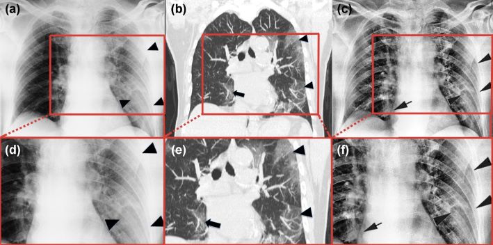
Immagine 1 - La figura mostra un esempio dell’applicazione di PACE, attraverso il confronto tra le seguenti immagini: (a) radiografia tradizionale, (b) immagine ottenuta con la TAC e (c) immagine radiografica elaborata con PACE. Grazie ad un miglioramento dei contrasti, è possibile riconoscere come l’elaborazione con PACE metta in evidenza un numero di lesioni polmonari, indicate con la freccia nera, non presenti nella radiografia tradizionale. Gli ingrandimenti (d, e, f) rendono ancora più visibile il confronto e la potenza del metodo, confermato dalle immagini TAC.
Picture #1 - The figure shows an example of the application of PACE, by comparing the following images: (a) traditional radiography, (b) image obtained with CT and (c) radiographic image processed with PACE. Thanks to an improvement in contrasts, it is possible to recognize how the processing with PACE highlights a number of lung lesions, indicated by the black arrow, not present in traditional radiography. The magnifications (d, e, f) make the comparison and the power of the method, confirmed by the CT images, even more visible